Cardio-MRI
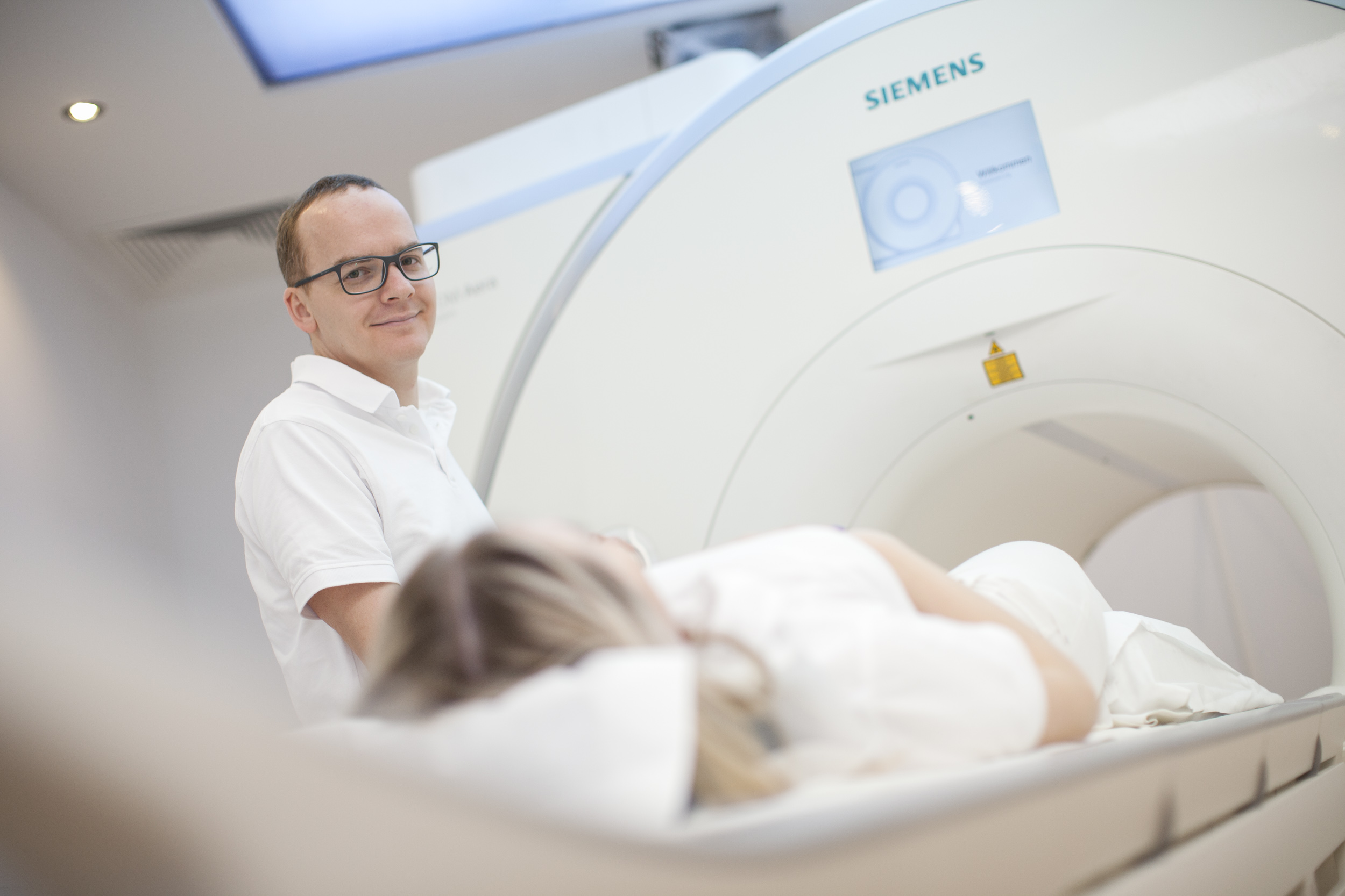
Magnetic Resonance Imaging is a pain-free layered imaging of the heart. It is free of radiation (no radio activity) and reconstructs an image of the beating heart.
This is what happens in a nutshell:
- the patient is placed in front of a magnet
- an electro-magnetic radio wave is emitted
- radio wave is switched off again
- patient sends a response signal which is recorded
- image reconstruction takes place
- constriction of coronary arteries (coronary heart condition, CHC)
- heart muscle inflammation (myocarditis chronic or acute)
- heart valve defect (e.g. leaky aortic valve)
- heart tumour or blood clot
- storage disease (iron storage disease)
- systemic disease (sarcoidosis) etc.
This is a procedure that is unrivalled, in other words, for many conditions there is no equivalent examination method.
Stress-MRI: While lying on the examination table, you are injected a medicine which leads to increased heart beat (similar to conditions under stress, comparable to physical exercise). The medication I use is either Adenosine or Dobutamine, depending, among other things, on possible previous illnesses, e.g. Asthma. This procedure requires an empty stomach (min. 6 hours prior to examination).
For a stress-MRI using Adenosine you are not allowed to consume coffee, tea or chocolate 24 hours prior to examination.
For a stress-MRI with Dobutamine no beta blockers must be used 48 hours prior to examination.
In case you are equipped with an MRI-compatible (MRI-conditional) cardiac pacemaker system (including probes) bad-quality images are to be expected, in this case I advise against a cardiac MRI. If you wear metal implants/prosthesis inform us prior to examination.
In any case, I will conduct a detailed consultation with you which answers all questions relating to the examination method.


